The embryo grading process assesses embryo quality and requires a vast amount of training and skill. Our embryologists use their experience to grade the embryos and select the best embryo for transfer. Please keep in mind that regardless of the grade, we only freeze and transfer embryos that we believe have a chance of resulting in a successful pregnancy.
Once fertilized, embryos will continue to develop in our laboratory for up to seven days. A blastocyst is the most advanced stage an embryo can achieve in culture and typically occurs 5 – 7 days post-fertilization. Only the best quality embryos develop to the blastocyst stage. A blastocyst has two distinct cell types and a central cavity filled with fluid:
Number describing overall expansion of the fluid cavity
Initially blastocysts are given a numerical score from 1 to 6 based upon their degree of expansion and hatching status. This part of the assessment can be performed on a dissection microscope.
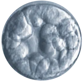
1 – Early Blastocyst
Blastocoel is less than half the volume of the embryo
2 – Blastocyst
Blastocoel is more than half the volume of the embryo
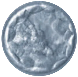
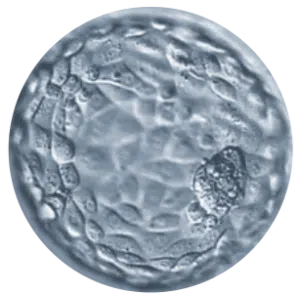
3 – Full Blastocyst
Blastocoel completely fills the embryo
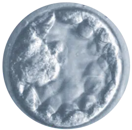
4 – Expanded Blastocyst
Blastocoel volume is now larger than that of the early embryo and the zona is thinning
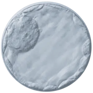
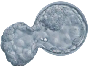
5 – Hatching
Embryo has breached the zona pellucida and is hatching out of its shell
6 – Hatched
Embryo is completely hatched
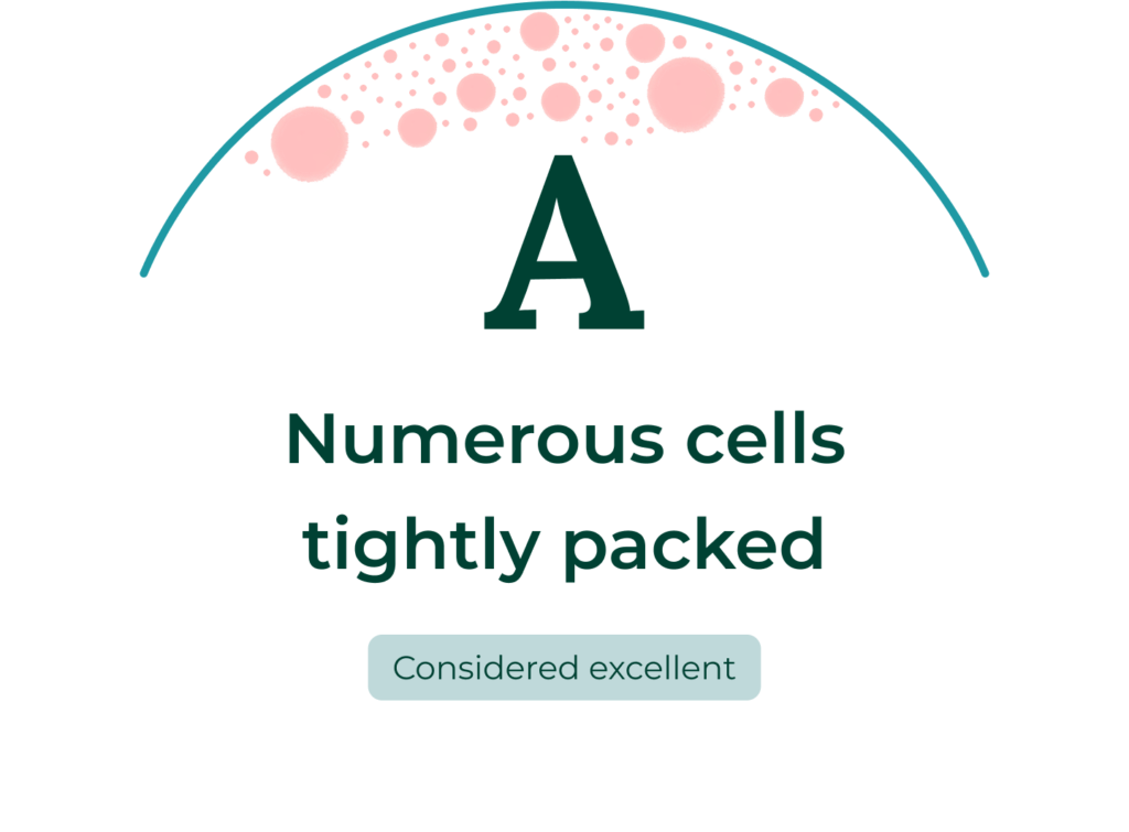
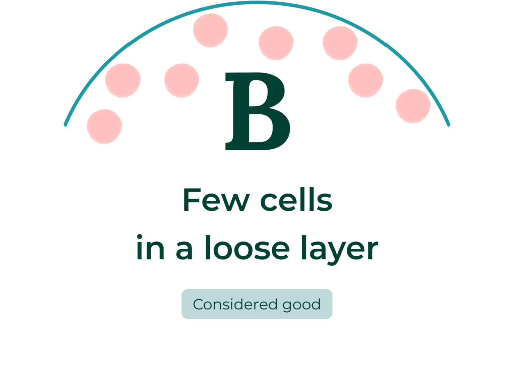
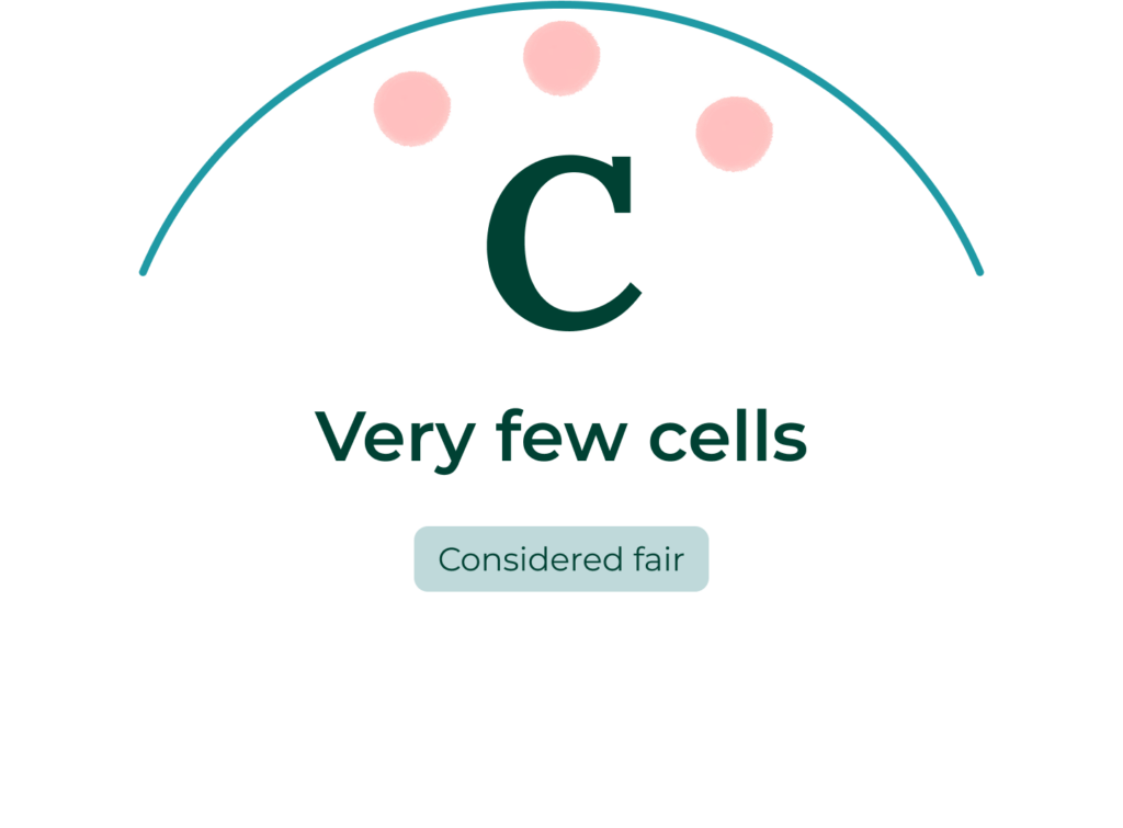
Letter grade for inner cell mass
The second step in scoring the blastocysts should be performed on an inverted microscope. The development of the inner cell mass (ICM) can then be assessed. The ICM is given a letter, the highest score being an A.
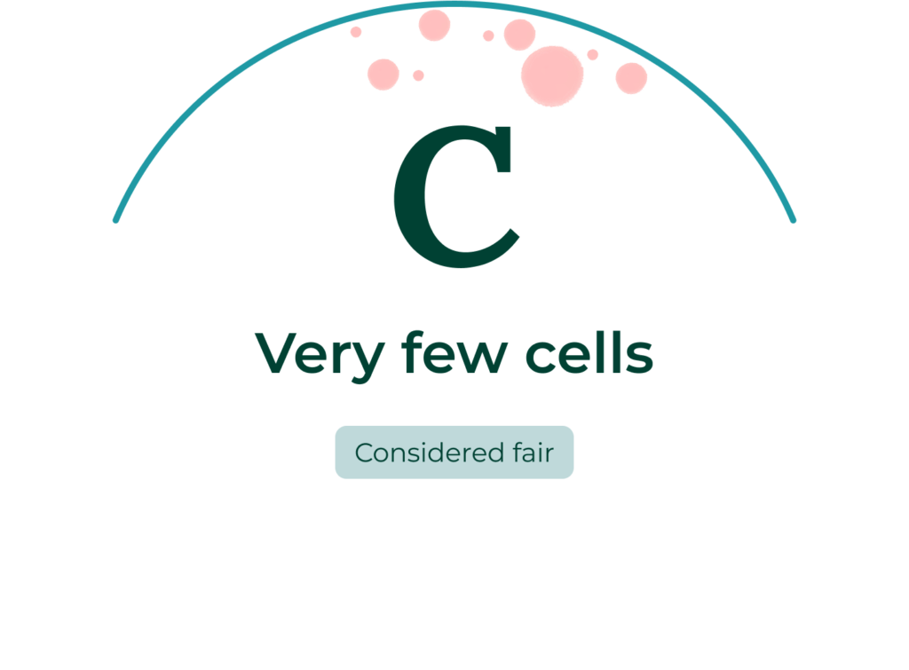
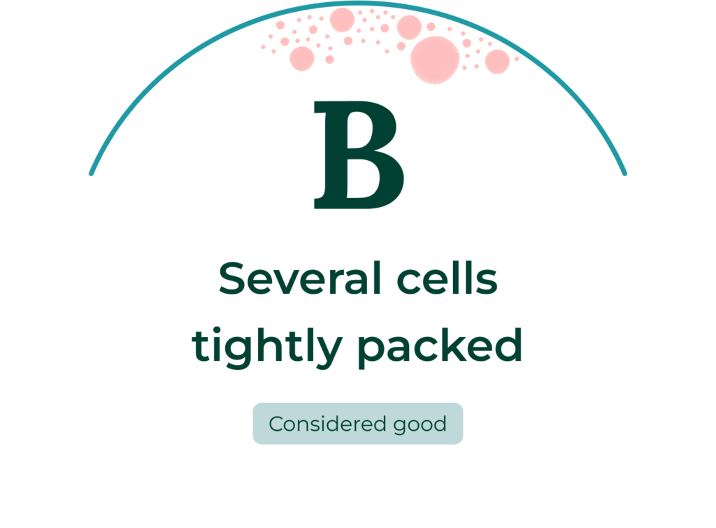
Letter grade for trophectoderm
The third step in scoring the blastocysts should be performed on an inverted microscope. The development of the trophectoderm can then be assessed. The trophecoderm is given a letter, the highest score being an A.
Learn more about our laboratory services
About our laboratory
Our laboratory team leads the way through research and innovation contributing to nationally recognized success rates.
EmbryoScope+
Time-lapse technology enables CCRM Fertility’s IVF experts and patients to view embryo development from its first cells.
PGT-A
Preimplantation genetic testing for aneuploidies (PGT-A) is a tool used to optimize the efficiency of IVF, increasing the likelihood of a successful pregnancy.
PGT-M and PGT-SR
Preimplantation genetic testing for monogenic/single gene defects (PGT-M) and (PGT-SR) are tests that help evaluate embryos for genetic risk factors.
Semen analysis
A semen analysis is performed in order to assess the viability of sperm before treatment at CCRM.
Additional embryo assessment tools
EmbryoScope+
Embryo grading is much more than the embryos’ isolated appearance. It also includes their morphokinetic development as witnessed via the EmbryoScope+.
Preimplantation genetic testing for aneuploidies (PGT-A, formerly known as PGS)
The chromosomal status of an embryo can be determined with preimplantation genetic screening which evaluates all 46 chromosomes within a few cells carefully removed from each embryo. PGT-A provides information regarding the chromosomal health of the embryo.
IVF For ME (Maximal Efficiency)
EmbryoScope+ time-lapse technology, when used in combination with PGT-A is a powerful tool in the decision-making process. This is why our IVF For ME program includes PGT-A and the use of EmbryoScope+ to monitor embryo development from its first cells.